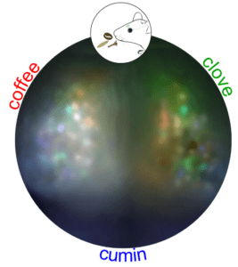
Exploring Endogenous Signals in Mouse Brain Imaging
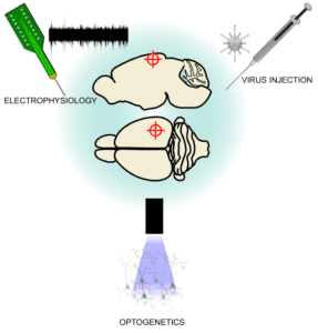
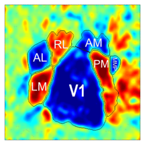
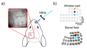
Autofluorescence imaging (AFI) is another, perhaps less commonly known, technique used to measure neuronal activity. It relies on the detection of changes in fluorescence from endogenous mitochondrial proteins inside neurons, named flavoproteins. During aerobic energy metabolism, these flavoproteins are oxidized which is translated by an increase in the molecule’s autofluorescence. Therefore, this signal can be used to measure neuronal activity given that an increase in neuronal responses is directly linked to an increase in its metabolic rate. In this blog, we explore the mouse primary somatosensory cortex function using both intrinsic and autofluorescence imaging techniques.